Glaucoma Explained: What To Know And Managing It Effectively

Introduction To Glaucoma

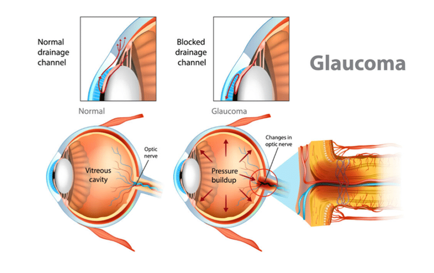
Why Do People Get Glaucoma

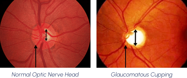
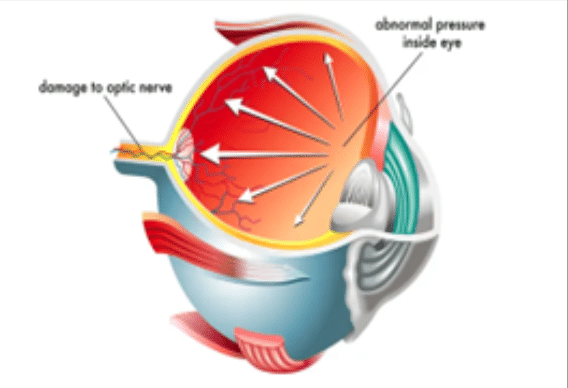
Glaucoma, often referred to as the “silent thief of sight”, can progress without noticeable symptoms until significant vision loss has occurred, making it the leading cause of blindness worldwide.

Understanding glaucoma is crucial as it silently progresses, often leading to significant and irreversible vision loss. If glaucoma goes undiagnosed, it can gradually and irrevocably deteriorate vision.

Introduction To Glaucoma

Risk Factors of Glaucoma

Being aware of these risk factors can help individuals take proactive steps towards monitoring their eye health. Regular eye examinations and consultations with an eye care professional are crucial for those at higher risk, ensuring early detection and effective management of glaucoma, which can damage the optic nerve. Here are some key risk factors to consider:

1. Age

The risk of developing glaucoma increases with age, particularly for individuals over the age of 60. Children can also be affected by congenital glaucoma, which presents early in life, This form is rare but underscores the spectrum of ages impacted by this condition.

2. Family History

A family history of glaucoma significantly raises the risk, suggesting a genetic predisposition to the condition.

3. Ethnicity

Certain ethnic groups, such as people of African, Asian, and Hispanicdescent, are at a higher risk of developing glaucoma.

4. Medical Conditions

Conditions such as diabetes, hypertension, and heart disease can increase the risk of glaucoma.

5. High Intraocular Pressure

Elevated intraocular pressure (IOP) is one of the primary risk factors for glaucoma. Regular eye exams can help monitor and manage IOP levels.

6. Thin Corneas

Individuals with thin corneas are at a higher risk of developing glaucoma, as this can affect the accuracy of IOP measurements.

7. Eye injuries


Eye injuries
Previous eye injuries or trauma can lead to secondary glaucoma, increasing the risk of the condition.

Eye Structures That Affect Glaucoma: A Layman’s Explanation

Glaucoma is a condition that can lead to vision loss, and it involves several key parts of the eye. Understanding these parts of the eye can help you grasp how glaucoma develops.

  • 1. Optic Nerve

    • What It Does: Think of the optic nerve as a cable that sends visual information from your eye to your brain.
    • How It Affects Glaucoma: When glaucoma damages this “cable,” it can lead to vision loss
  • 2. Intraocular Pressure (IOP)

    • What It Does: This is the pressure inside your eye, similar to how air pressure keeps a balloon inflated.
    • How It Affects Glaucoma: High eye pressure can damage the optic nerve, leading to glaucoma.
  • 3. Anterior Chamber

    • What It Does: This is the front part of your eye, filled with a clear fluid that nourishes the eye.
    • How It Affects Glaucoma: If the fluid doesn’t drain properly, pressure can build up, causing glaucoma.
  • 4. Trabecular Meshwork

    • What It Does: This is like a drain in your eye that helps fluid flow out.
    • How It Affects Glaucoma: If this drain gets clogged, fluid can’t leave the eye, increasing pressure.
  • 5. Canal of Schlemm

    • What It Does: This is a tiny channel that carries fluid from the trabecular meshwork out of the eye.
    • How It Affects Glaucoma: If this channel is blocked, fluid builds up, raising eye pressure.
  • 6. Iris

    • What It Does: This is the coloured part of your eye that controls the size of your pupil.
    • How It Affects Glaucoma: In some types of glaucoma, the iris can block the drainage angle, causing a sudden increase in pressure.
  • 7. Ciliary Body

    • What It Does: This part of the eye produces the fluid that fills the anterior chamber.
    • How It Affects Glaucoma: If too much fluid is produced or not enough drains out, pressure can rise.
  • 8. Cornea

    • What It Does: This is the clear, front surface of your eye that helps focus light.
    • How It Affects Glaucoma: The thickness and shape of the cornea can affect pressure readings and glaucoma risk.
  • 9. Retina

    • What It Does: This is the layer at the back of your eye that senses light and sends signals to the brain.
    • How It Affects Glaucoma: Damage to the optic nerve can lead to loss of retinal cells, affecting vision.

Signs & Symptoms of Glaucoma (Asymptomatic)

Recognizing the early signs of glaucoma is crucial for timely intervention and preserving
vision. Given that early detection and treatment often begin only after significant vision loss has occurred.

1. Early Stage Glaucoma

Gradual Loss of Peripheral Vision: One of the earliest signs of glaucoma is the gradual loss of peripheral vision. This often goes unnoticed (asymptomatic) until significant damage has occurred

2. Mid To Late Stage Glaucoma

Tunnel Vision: As glaucoma progresses, individuals may experience tunnel vision, where the field of vision narrows, making it difficult to see objects on the periphery.

Types of Symptomatic Glaucoma

Glaucoma encompasses several types, each with distinct symptoms and characteristics. Understanding these types can aid in early detection and effective management. Here are the primary types of symptomatic glaucoma:

1. Congenital Glaucoma

Symptoms: In infants, symptoms include enlarged eyes, cloudiness of the cornea, excessive tearing, and sensitivity to light.
Characteristics: This rare type is present at birth and results from abnormal development of the eye’s drainage system.

2. Pigmentary Glaucoma

Symptoms: Blurred vision, halos around lights, and occasional eye pain, often exacerbated by physical activity.

Characteristics: Pigment granules from the iris can flake off and clog the drainage channels cause increased IOP, resulting as a secondary glaucoma.

3. Exfoliative Glaucoma

Symptoms: Similar to POAG, with gradual vision loss and potential for more rapid progression.
Characteristics: Flaky, dandruff-like material peels off the outer layer of the lens and clogs the drainage system.

4. Eye Injuries

Description: Trauma to the eye, such as blunt force or penetrating injuries, can damage the eye’s drainage system, leading to increased intraocular pressure (IOP).

Impact: This can result in secondary glaucoma, often requiring prompt medical attention to prevent vision loss.

5. Inflammation

Description: Conditions like uveitis, which cause inflammation inside the eye, can block the drainage channels or lead to the formation of scar tissue.

Impact: This blockage can increase IOP, resulting in secondary glaucoma.

6. Medications

Description: Long-term use of certain medications, particularly corticosteroids, can raise IOP.

Impact: This can lead to the development of secondary glaucoma, necessitating regular monitoring and possible adjustment of medication.

7. Eye Surgery

Description: Complications from eye surgeries, such as cataract removal, can sometimes lead to secondary glaucoma.
Impact: Post-surgical changes in the eye’s anatomy can affect fluid drainage, increasing IOP.

8. Other Eye Conditions

Description: Conditions like advanced cataracts, retinal detachment, or tumours can obstruct the eye’s drainage system.
Impact: These obstructions can lead to secondary glaucoma.

Can Glaucoma Be Treated?

Glaucoma, a complex eye condition, can indeed be managed with effective treatment strategies tailored to the individual’s specific needs.

Treatments for Glaucoma: An Overview

Eyedrops

Firstly, eye drops play a significant role in reducing intraocular pressure, often being the first line of defence. These medications are designed to either decrease the production of fluid within the eye or enhance its drainage, thereby alleviating the pressure that could lead to optic nerve damage.

Here are some common medical eyedrop treatments for glaucoma:

1. Prostaglandin Analogues

Function: Increase the outflow of aqueous humour, reducing IOP.

Examples: Latanoprost, Bimatoprost, Travoprost.

Usage: Typically administered as eye drops, usually once daily.

2. Beta Blockers

Function: Decrease the production of aqueous humour.
Examples: Timolol, Betaxolol.
Usage: Administered as eye drops, usually once or twice daily

3. Alpha Agonists:

Function: Reduce aqueous humour production and increase outflow.
Examples: Brimonidine, Apraclonidine.
Usage: Administered as eye drops, usually twice or three times daily.

4. Carbonic Anhydrase Inhibitors:

Function: Decrease the production of aqueous humour.
Examples: Dorzolamide, Brinzolamide.
Usage: Available as eye drops or oral medications, typically used two to three timesdaily.

5. Rho Kinase Inhibitors:

Function: Increase the outflow of aqueous humour through the trabecular meshwork.
Examples: Netarsudil.
Usage: Administered as eye drops, usually once daily.

6. Combination Medications:

Function: Combine two different types of medications to enhance effectiveness.
Examples: Timolol/Dorzolamide, Brimonidine/Timolol.
Usage: Administered as eye drops, typically used twice daily.

Laser Therapy

Laser treatment is another promising approach, particularly useful when medications alone are not sufficient. This procedure is minimally invasive, utilising a focused beam of light to improve fluid drainage or reduce fluid production, which helps in controlling the overall pressure within the eye.

Here are some common laser procedures for glaucoma and what to expect during recovery:

1. Laser Trabeculoplasty (LT)

Function: Uses laser energy to improve drainage through the trabecular meshwork.

Usage: Often used for open-angle glaucoma, can be repeated if necessary.

Procedure: A small flap is created in the sclera (white part of the eye), and a reservoir (bleb) is formed under the conjunctiva (the outer covering of the eye) to allow aqueous humour to drain out of the eye, reducing IOP.

Recovery: Patients may experience blurred vision and discomfort for a few weeks. Anti-inflammatory and antibiotic eye drops are typically prescribed to prevent infection and control inflammation. Regular follow-up visits are crucial to monitor healing and IOP levels.

2. Laser Peripheral Iridotomy

Function: Creates a small hole in the iris to improve fluid flow.

Usage: Commonly used for angle-closure glaucoma

Procedure: A laser creates a small hole in the iris to improve fluid flowbetween the anterior and posterior chambers of the eye, reducing IOP.

Recovery: Patients may experience mild discomfort and sensitivity to light for a few days. Anti-inflammatory eye drops are often prescribed. Follow-up visits are necessary to monitor IOP and ensure the iridotomy remains open.

Other Surgical Interventions

More advanced cases may necessitate surgical interventions to create new drainage pathways. While surgery carries inherent risks, modern techniques have improved the overall safety and efficacy. With vigilant monitoring and adherence to treatment plans, individuals with glaucoma can maintain their vision and lead fulfilling lives.

Here are some common surgical procedures for glaucoma and what to expect during recovery:

1. Minimally Invasive Glaucoma Surgery (MIGS)

Function: Involves smaller incisions and aims to reduce IOP with fewer complications.

Examples: iStent, Xen Gel Stent.

Usage: Often used in conjunction with cataract surgery.

Recovery: Recovery is generally quicker and less painful compared to traditional surgeries. Patients may experience mild discomfort and blurred vision for a few days. Eye drops are prescribed to aid healing and prevent infection. Regular follow-ups are essential to monitor IOP and ensure the
stent is functioning properly.


2. Cyclophotocoagulation

Function: Uses laser to reduce the ciliary body’s ability to produce aqueous humour.

Usage: Typically reserved for advanced or refractory glaucoma.

Procedure: A laser is used to reduce the ciliary body’s ability to produce aqueous humour, thereby lowering IOP. This can be done externally (transscleral) or internally (endoscopic).

Recovery: Patients may experience discomfort, redness, and blurred vision for a few days to weeks. Anti-inflammatory and antibiotic eye drops are prescribed. Regular follow-ups are crucial to monitor IOP and assess the effectiveness of the procedure.

3. Glaucoma drainage devices

Function: Involves surgically implanting a small drainage device to facilitate the outflow of aqueous fluid from the eye, helping to lower intraocular pressure (IOP).

Usage: Ahmed Valve, Baerveldt Implant.

Procedure: A small tube is inserted into the eye to divert fluid to a reservoir placed under the conjunctiva (the outer layer of the eye), allowing the fluid to be absorbed naturally by surrounding tissue.

Recovery: Patients may experience redness, irritation, or blurred vision for several days to weeks. Anti-inflammatory and antibiotic eye drops are prescribed to prevent infection and control inflammation. Regular follow-up appointments are essential to monitor eye pressure and ensure proper functioning of the device.

4. Combined Cataract and Glaucoma Surgery

Function: Simultaneously addresses cataracts and glaucoma by removing the cataract and improving fluid drainage from the eye to lower IOP.

Usage: Recommended for patients who have both visually significant cataracts and glaucoma.

Procedure: Cataract surgery (phacoemulsification) is combined with a glaucoma procedure such as trabeculectomy or MIGS. The removal of the cataract can open the drainage angle, promoting better outflow of aqueous fluid and reducing eye pressure.

Recovery: Recovery is similar to standard cataract surgery, with some added monitoring for IOP. Mild discomfort, light sensitivity, and blurry vision are common in the first few days. Eye drops are prescribed to support healing and manage inflammation. Follow-up visits are crucial to ensure both surgical components are successful and that eye pressure is well-controlled.

Aftercare and Recovery Tips

1. Follow Medication Instructions

Use prescribed eye drops and medications as directed to prevent infection and control inflammation.

2. Avoid Strenuous Activities

Refrain from heavy lifting, bending over, or strenuous activities that could increase IOP.

3. Protect Your Eyes

Wear protective eyewear as recommended by your doctor, especially when outdoors or in dusty environments.

4. Attend Follow-Up Appointments

Regular check-ups are crucial to monitor IOP, ensure proper healing, and adjust treatment if necessary.

5. Report Any Issues

Contact your eye care professional immediately if you experience severe pain, sudden vision changes, or signs of infection.

Note icon
The symptoms and possible related eye conditions/diseases in this section are for general reference only, and do not contain all visual symptoms or all possible related conditions or diseases. If you have any unusual vision symptoms, speak with your ophthalmologist.

At Hesed Eye Specialists, we are dedicated to not just restoring, but truly enhancing your vision.

Trust in our expertise and commitment to excellence as we accompany you on your journey towards clearer, brighter, and healthier eyes. Experience the profound difference that an eye clinic with unparalleled technology and compassion can make in your life and vision. 

Book An Appointment
Make An Appointment

Free Shuttle Services

There are free shuttle services available on Monday to Friday from 8.30am to 5.10pm and Saturday from 8.30am to 1pm.*

* These services are not available on Sundays and public holidays. Please note that pick-up and drop-off are at the designated stops only and the schedule is subject to change due to traffic conditions and other factors without prior notice.

Mondays to Fridays (Excluding Public Holidays):

Gleneagles Hospital - Mount Elizabeth Hospital - The Heeren

Gleneagles Hospital
Mount Elizabeth Hospital
The Heeren
8.30 am
8.30 am
8.30 am
8.30 am
8.30 am
8.30 am
8.32 am
8.32 am
8.32 am
8.30 am
8.32 am
8.32 am

Saturdays (Excluding Public Holidays):

Gleneagles Hospital - Mount Elizabeth Hospital - The Heeren - Mount Elizabeth Novena

Gleneagles Hospital
Mount Elizabeth Hospital
The Heeren
Mount Elizabeth Novena Hospital
8.30 am
8.30 am
8.30 am
8.32 am
8.30 am
8.30 am
8.30 am
8.30 am
8.32 am
8.32 am
8.32 am
8.32 am
8.32 am
8.32 am
8.32 am
8.30 am

For faster response, please Whatsapp us