Understanding Retinal Health and Eye Floaters

Introduction To Floaters

Have you ever paused to wonder how floaters affect your vision?

Imagine them as tiny dancers, suspended in the grand ballroom of your eye, occasionally drifting across your field of view, much like eye floaters. While they may pique curiosity, it is essential to understand that, more often than not, they are harmless companions on our visual journey, subtly reminding us of the intricate complexity and wonder of our retinal world.

What Are Floaters?

Eye floaters are small entities that drift across your field of vision, usually appearing as specks, dots, or lines, potentially leading to vision problems.

They originate from the natural ageing of the eye’s vitreous, a gel-like substance that fills most of the eye and provides structural support.

As the vitreous ages, it transitions from a gel into a more liquid form, causing tiny strands to form, and diabetes can sometimes accelerate this process. These strands cast shadows on the retina, resulting in the appearance of floaters.

Though generally benign, these visual phenomena can occasionally signal a more serious issue if new floaters suddenly appear or increase dramatically.

In such instances, it is crucial to consult a specialist to rule out potential retinal concerns.

Causes of Retinal Floaters

Retinal floaters primarily arise from the natural aging process of the eye’s vitreous. As we get older, the vitreous gel that maintains the eye’s shape begins to shrink and liquefy.

Other factors that may contribute to floaters include eye injuries, inflammation, or uveitis.

The gradual detachment of the vitreous from the retina, known as posterior vitreous detachment, can cause fine, fibrous strands to develop, which are perceived as floaters. Myopia, or short-sightedness, is one of the common risk factors, alongside previous eye surgeries such as cataract operations.

Moreover, trauma and retinal tears significantly increase the likelihood of floaters in the visual field. It is prudent to remain vigilant, as any sudden changes or increase in floaters could indicate a more pressing retinal condition that necessitates immediate medical attention. By understanding these causes, individuals can better appreciate the complexities of their eye health and act decisively in safeguarding their vision.

Recognising Symptoms of Floaters

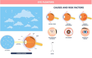
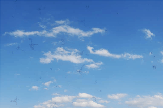
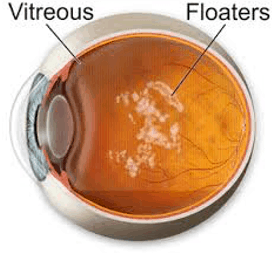
Floaters, those intriguing specks or cobweb-like shadows drifting across one’s vision, signify natural changes within the vitreous gel of the eye. Far from being a mere distraction, these floaters serve as reminders of the intricate and marvellous workings of the human eye.

Often appearing as dots, lines, or ambiguous shapes, these visual phenomena can arise suddenly, though they might have been building up subtly over time. Their presence in our field of vision, while typically benign, commands our awareness of any sudden onsets or dramatic increases in their number.

Moreover, floaters frequently accompany flashes, bursts of light seen even in darkness. Such flashes, stemming from mechanical pulls on the retina, can heighten one’s intuition that something may warrant medical investigation.

It’s vital to bear in mind that floaters naturally evolve with age, predominantly benign and stable in nature, requiring no treatment unless dramatic changes surface. Awareness and observation become key, as monitoring one’s symptoms can vastly aid in distinguishing between harmless floaters and more significant retinal issues.

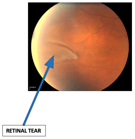
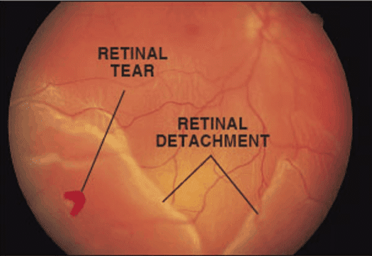
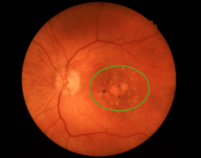
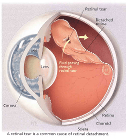
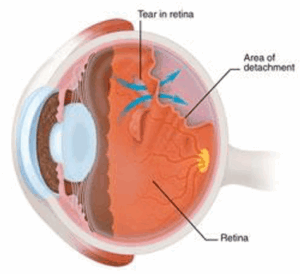
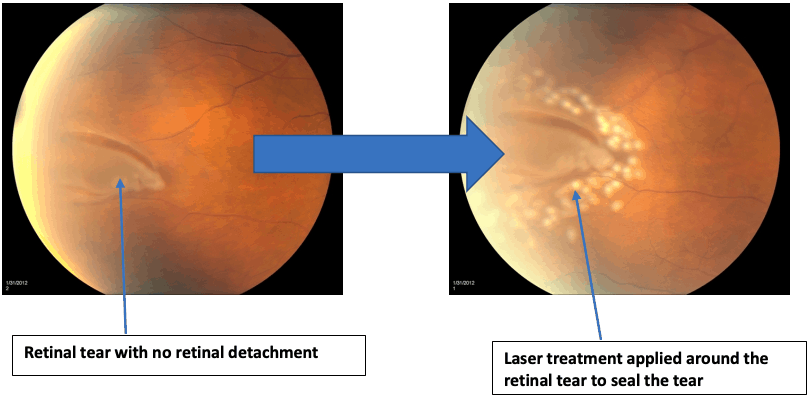
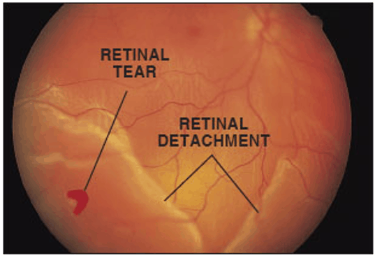
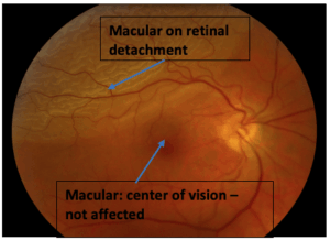
When new floaters emerge or an unexpected ‘shower’ of these spots occurs, it may be indicative of more serious retinal conditions such as a tear or detachment that requires urgent attention. Professional evaluation ensures peace of mind and can prevent potential complications from escalating.

Ultimately, the dance of floaters across one’s vision is a call to remain vigilant regarding eye health. By recognising these symptoms, individuals empower themselves, fortifying their commitment to an enduring and flourishing vision.


When to Seek Medical Attention

Experiencing new floaters or a sudden ‘shower’ of black spots could signal more serious retinal conditions, such as a tear or detachment. Immediate professional evaluation is crucial to mitigate potential complications and preserve your precious vision.

In addition to these alarming visual changes, if you notice an increase in flashes, a shadow obstructing your vision, or a grey curtain appearing in your field of view, consult an eye specialist urgently. These symptoms could indicate a retinal break or detachment, requiring swift medical intervention to protect and sustain your eye health. By promptly addressing such signs, you take a proactive step towards preserving the clarity and brilliance of your vision for years to come

1. New Onset of Floaters

Floaters might appear suddenly—whether dots or cobwebs—the visual spectacle often has a deeper story. Early detection of changes in your vision, such as floaters, is vital for preserving eye health.
A sudden appearance of floaters warrants immediate attention, as they may indicate underlying retinal issues. Acting promptly can prevent minor annoyances from escalating into more severe ocular complications.
Noticing changes in your vision—be it a shadow, flashes, or floaters—demands a vigilant and immediate response. Seeking professional guidance swiftly is the most effective strategy for maintaining optimal eye health.

2. Significant Increase in Floaters

An unexpected rise in floaters can be more than a mere nuisance—could it be a sign of underlying retinal concerns?
In many cases, an abrupt increase in floaters can serve as a forewarning of potential retinal tears. It is imperative to seek the expertise of an eye specialist immediately to diagnose the issue accurately.
Recognising the significance of a sudden influx of floaters can make all the difference in preserving your vision’s integrity. Delay not; take prompt action to ensure that your eye health remains uncompromised and vibrant.
Eye health is a precious asset; remain attentive and responsive to any alterations, no matter how small.

3. Persistent Light Flashes

Persistent light flashes often manifest as unexpected, fleeting bursts of illumination that may appear even in complete darkness.

  • New occurrence of light flashes
  • Sudden intensification in frequency or brightness
  • Visibility of flashes without external light
  • These phenomena may signal the vitreous gel’s mechanical pull on the retina.
    Immediate consultation with an eye care professional is advised to prevent serious retinal conditions.

    Understanding Retinal Tears

    A retinal tear occurs when the vitreous gel inside your eye pulls on the retina with sufficient force to create a break. This event can significantly impact your visual health.

    Retinal tears can predispose the eye to more serious conditions like retinal detachment.

    Occasionally, individuals might experience an abrupt increase in eye floaters or flashes due to tractional forces on the retina, often associated with posterior vitreous detachment.

    Should these symptoms manifest, seeking immediate ophthalmic evaluation is crucial to mitigate potential complications.

    Lasers applied to seal retinal tears can be life-changing, thwarting possible progression to retinal detachment, and preserving the quality of visual acuity.

    Remember, the power of your eyes is as remarkable as it is delicate. Early detection through vigilant attention to symptoms ensures a pathway to sustained eye health.

    Understanding Retinal Tears

    Symptoms of Retinal Tears

    A retinal tear requires swift attention to prevent the condition from advancing to more serious complications such as retinal detachment.

    The primary treatment modality for retinal tears is laser retinopexy, a technique that effectively seals the tear by promoting scar formation. This proactive intervention leverages advanced laser technology to safeguard ocular integrity and preserve visual acuity.

    By preventing further separation of the retina, laser retinopexy helps to avert the progression of the tear. This innovative solution is typically performed in a clinical setting, offering patients a convenient and minimally invasive treatment route.

    The procedure’s precision minimises the risk of complications and boasts a high success rate, reassuring patients of their visual future. While laser retinopexy is transformative, it underscores the necessity for early detection and immediate action upon identifying symptoms.

    Thus, if you experience any alarming symptoms, consulting an eye specialist can be your best decision.

    Symptoms of Retinal Detachment

    Understanding the symptoms of retinal detachment is essential, as it poses significant challenges to one’s vision, particularly when paired with vision problems, and may even threaten it in severe cases.

    Typically, the initial signs manifest as sudden bursts of floaters, flashes of light, or symptoms of uveitis.

    Some individuals, particularly those with conditions like diabetes, encounter dark shadows or a “curtain” effect sweeping across part of their vision, indicating that the retina may be peeling away from its potentially life-sustaining layer.

    Prompt medical evaluation for these symptoms is crucial. If tackled early, there is a greater probability of reversing the condition through appropriate intervention. Our eyes entrust us with the world’s visual wonders; thus recognising any abnormalities and acting swiftly can save one’s sight and preserve life’s beauty.

    Symptoms of Retinal Detachment

    Treatment for Retinal Detachment

    Treating retinal detachment requires timely intervention to preserve vision and prevent further complications, and each case requires a tailored approach depending on its severity

    Thorough scans and tests determine the most effective treatment option.

    Scleral buckling, a surgical intervention to counteract the force causing detachment, often involves placing a silicone band around the eye. This procedure, complemented with fluid drainage, encourages the retina to reattach securely, ensuring robust visual health.

    Another advanced solution, vitrectomy, removes the vitreous gel tugging at the retina. Surgeons might insert a gas or oil bubble to stabilise the retina during the healing phase. While the introduction of the gas bubble necessitates avoiding air travel temporarily, such small sacrifices are a “worth-it” exchange for healthy eyesight.


    Risks of Retinal Detachment Surgery

    Retinal detachment surgery carries potential risks.

    While the procedure is often successful, it does not come without challenges. The inherent complexities of eye surgery mean there are risks associated with anaesthesia, potential infections, and the surgery itself. Infection rates are minimal thanks to sterilisation practices, but like any operation, a small infection risk remains.

    The risk of bleeding is low.

    There is a slight chance of increased intraocular pressure, potentially leading to glaucoma. The incidence of this is rare but may require further treatment through eye drops or additional surgery to manage the condition effectively.

    Finally, there is a possibility of cataract formation and eye floaters post-operation, particularly amongst older patients. However, cataract development can be expertly addressed with subsequent surgery, ensuring the best possible outcome. Equipped with advanced technologies and guided by skilled surgeons, today’s procedures generally ensure a high success rate and safe recovery.

    What Is an Epiretinal Membrane?

    An epiretinal membrane is a thin, fibrous layer forming on the surface of the retina, particularly affecting the macula, which is critical for sharp central vision.

    This condition can lead to visual disturbances, requiring expert evaluation.

    Such membranes can arise from various situations, including previous retinal tears, detachments, or vascular conditions, such as diabetes-related retinopathy or vein occlusion. While the presence of an epiretinal membrane can affect vision, many individuals may remain symptom-free.

    The development of an epiretinal membrane is frequently associated with the natural ageing process and the accompanying changes in the eye’s vitreous gel. If visual impairment occurs or worsens, surgical intervention may be considered necessary. Advanced surgical techniques, employed by skilled ophthalmologists, can often restore or improve vision, ensuring that those affected can continue to engage fully in their daily pursuits.

    What Is an Epiretinal Membrane?

    Causes of Epiretinal Membrane

    Understanding the causes behind the formation of an epiretinal membrane is pivotal for early diagnosis and effective management.

    Primarily, an epiretinal membrane can develop as a consequence of retinal changes and stress factors, such as retinal tears and detachments, which disrupt the retinal surface. Retinal vascular diseases including diabetic retinopathy or vein occlusion also contribute to its development by prompting an inflammatory process, such as uveitis, or initiating cellular reactions that result in membrane formation.

    Interestingly, age plays a significant role as a natural precursor to these membranes. As individuals age, changes in the vitreous gel of the eye instigate tiny tractional forces on the retina, sometimes contributing to conditions like posterior vitreous detachment, leading to the potential formation of a thin, fibrous layer on the macula’s surface.

    Furthermore, surgical procedures related to eye conditions, particularly those addressing retinal issues, may inadvertently trigger epiretinal membrane growth. While these occurrences are relatively rare, understanding their origins is crucial in identifying at-risk individuals and advocating timely assessments, ensuring that vision complications do not become debilitating and life-limiting.

    Treatment Options for Epiretinal Membranes

    Epiretinal membranes, while often stable, sometimes require surgical intervention when vision is significantly affected and other treatments are ineffective.

    Primarily, vitrectomy stands at the forefront as a preferred surgical approach. In this intricate procedure, the vitreous gel is delicately removed from the eye, providing the surgeon access to meticulously peel away the problematic epiretinal membrane. This procedure, tailored to reduce distortion, aims to enhance visual clarity and patient quality of life.

    Remarkably, the timing and nature of surgical intervention are pivotal in achieving optimal outcomes. For cases induced by retinal detachment or vascular issues, results may vary. Even with meticulous surgical techniques, certain factors can influence the prognosis and recovery time, emphasising the importance of early detection.

    In collaboration, advancements in surgical methodologies promise increasingly refined results, empowering individuals to confront vision challenges with tenacity. Innovations such as smaller gauge instruments and advanced imaging technologies elevate the precision of membrane removal, fostering a hopeful outlook for patients. Remaining informed and proactive ensures successful navigation through the complexities of epiretinal treatments.

    Understanding Macular Holes

    In the intricate landscape of retinal health, macular holes present as an intriguing phenomenon affecting the eye’s central vision. These tiny tears or openings in the macula can lead to blurred or distorted central vision, impacting daily activities such as reading and recognising faces. As age advances, the risk of developing a macular hole increases, making awareness and timely intervention crucial.

    Understanding Macular Holes

    Emerging from the natural shrinkage of the vitreous gel, macular holes occur when this gel exerts traction on the retina. This action may result in openings that disrupt the fluid surface of the macula, altering vision. Detecting symptoms early can make a significant difference, as advanced cases may cause dark or blind spots in one’s central field of vision, escalating concerns.

    Treatment for macular holes often involves a surgical procedure known as a vitrectomy, where the vitreous gel is removed and a gas bubble is introduced. This method encourages the hole to close, and the macula to heal effectively. Patients are advised to maintain a face-down position to help the bubble press against the hole, facilitating closure over time.

    Despite the challenges, advances in surgical techniques and understanding of ocular health promise optimistic outcomes for those affected. Groundbreaking innovations in eye care ensure that interventions are precise and recovery is facilitated, supporting improved visual acuity post-surgery. The proactive management of eye health underscores a more hopeful future for individuals diagnosed with macular holes.

    With vigilance and early detection, solutions for macular holes continue to develop, offering reassurance and improving quality of life for many.

    Causes and Treatment of Macular Holes

    Macular holes are primarily caused by the natural ageing process, where vitreous shrinkage leads to traction on the retina

    As the vitreous gel within the eye diminishes with age, it can pull on the macula, causing a hole to form, and may also result in eye floaters. In some cases, this can result in blurry or distorted central vision, often accompanied by a troubling dark spot.

    Diagnosing a macular hole early can significantly enhance treatment outcomes. While the condition primarily afflicts individuals beyond the age of 60, regular eye examinations can facilitate timely detection and intervention.

    Treatment usually involves a surgical procedure called a vitrectomy, where the vitreous gel is removed, and a gas bubble is injected. This procedure promotes the natural healing process, allowing the macula to regain its function.

    Innovative treatments continue to evolve, offering brighter prospects and improved results for those challenged by macular holes.

    Note icon
    The symptoms and possible related eye conditions/diseases in this section are for general reference only, and do not contain all visual symptoms or all possible related conditions or diseases. If you have any unusual vision symptoms, speak with your ophthalmologist.

    At Hesed Eye Specialists, we are dedicated to not just restoring, but truly enhancing your vision.

    Trust in our expertise and commitment to excellence as we accompany you on your journey towards clearer, brighter, and healthier eyes. Experience the profound difference that an eye clinic with unparalleled technology and compassion can make in your life and vision. 

    Book An Appointment
    Floaters

    Quick Facts: Flashes & Floaters

    Early symptoms: 

    Floaters –Small specks, dots, circles, lines or cobwebs

    Flashes – Flashing lights or lightning, often noticed in the corner of vision, even in dark places

    Later symptoms:

    Retinal Break – sudden increase in black spots like a “shower” of black dots

    Retinal Detachment – a “curtain” or shadow blocking part of your vision

    Risk factors:

    Myopia (short-sightedness), Family history of retinal detachment, History of serious eye injury, previous eye surgery such as cataract surgery

    Treatment:

    Treatment is not usually needed for floaters. Floaters do not disappear as they are part of the eye, but become less noticeable with time.

    What are floaters?

    The first thing to know is that floaters are part of our eye.

    • They are formed from the vitreous gel that occupies 80 percent of the eye.
    • The vitreous gel shrinks and also changes from being gel-like to being more watery and stringy over time.This causes the formation of strands which cast tiny shadow on the retina which we see as floaters.
    • The vitreous gel is also attached to the retina initially. However, as it shrinks, it pulls away and detaches from the retina.
    • This may not occur completely, leaving parts of the vitreous gel still attached to parts of the retina.
    • When the attached parts of the vitreous gel pulls on the retina, we notice flashes in our eye. These flashes are not caused by stimulation from light entering the eye, but do to mechanical pull by the vitreous gel on the retina. Hence, flashes can occur in the dark without any surrounding light.

    When should you be worried about floaters and flashes?

    Floaters and flashes are usually not a worry. However, there are times when you should be seen by an eye doctor urgently:

    • New onset of floaters
    • Sudden increase in floaters
    • Increased flashing
    • Seeing a shadow blocking parts of your vision
    • Seeing a gray curtain covering parts of your vision

    These symptoms can suggest either a retinal break or a retinal detachment.

    Retinal Tears & Retinal Detachments

    What are Retinal Tears?

    The retina is a sheet of light-sensitive cells responsible for receiving light signals so we can see. The retina is attached to the inner wall of the eye.

    The vitreous gel is a gel-like substance that is attached to the retina. Over time, the vitreous gel shrinks and becomes watery.

     As the vitreous gel shrinks, it can pull on the retina causing a retinal tear.

    What is the symptoms for Retinal Tears?

    Symptoms of a retinal tear are similar to floaters. That is why it is important to have your floaters checked to rule out retinal tears. Retinal tears can have symptoms such as:

    • Sudden increase in number of floaters
    • Sudden appearance of flashes

    What is the treatment for Retinal Tears?

    In the case above, we have a retinal tear with no retinal detachment. The treatment in this case would be to perform laser retinopexy which is a clinic-based laser.

    In a laser retinopexy, laser is applied all around the tear. This helps to seal the tear and prevent vitreous from entering through the tear and detaching the retina.

    This clinic-based laser can only be performed if there Is no retinal detachment. If there is retinal detachment, surgery will be needed. So it is important to find and treat retinal tears early before they become retinal detachments.

    What are retinal detachments?

    Retinal detachments are when the retina detaches from the inner wall of the eye. This occurs when retinal tears are not sealed early. This results in fluid traveling through the retinal tear and under the retina causing a detachment.

    What are the symptoms of Retinal Detachment?

    Symptoms of a retinal detachment include:

    • Seeing a shadow in the side of your vision
    • Seeing a gray curtain moving across your vision
    • Sudden decrease in vision

    What is the treatment for Retinal Detachment?

     The best results from treatment occur when the retinal detachment has not affected the center of your vision. This usually means that the retinal detachment is not involving the central part of retina called the macular. This is called a macular on retinal detachment and will have better chance of recovering good vision after treatment. Macular off retinal detachment on the other hand, will not do as well, even after successful surgery. That is why it is critical to identify and treat early.

    Sclera Buckle

    This involves placing a band around the eye to counteract the force that is causing the retina to be detached. This often also involves drainage of fluid from under the detached retina, allowing reattach ent of the retina. This is surgical procedure that is carried out in the operating theatre.

    Vitrectomy

    This surgery involves removal of the vitreous gel that is pulling on the retina. Often a gas bubble may be placed in the eye to keep the retina attached while healing takes place. When a gas bubble is introduced, you may need to keep your head in a certain position for a time. Also you will not be able to fly for a period of time as the high altitudes can cause dangerous increase in eye pressure from expansion of the gas bubble. Over time, the gas bubble will gradually be replaced by your own body’s fluids.

    Sometimes, an oil bubble may be introduced instead of a gas bubble. An oil bubble will need to be removed later when the retina has fully attached.

    A vitrectomy can be combined with cataract surgery and/or a scleral buckle procedure.

    What are the risks of a vitrectomy surgery?

    Vitrectomy surgery has advanced in recent years and is a relatively safe procedure.  The risks include:

    • Infection: 1 in 2,000 chance
    • Retinal detachment: 1 in 100 chance
    • Bleeding in the eye: very rare
    • Glaucoma where the eye pressure become high: but usually this responds to treatment with eye drops
    • Cataract where the lens become cloudy: this is readily treatable
    Epiretinal Membrane

    What is an epiretinal membrane?

    An epiretinal membrane is a thin sheet of fibrous tissue that develops on the surface of the macular and can cause problems with central vision.

     This is because this membrane can contract and cause distortion of the retina. As the macular is responsible for visually demanding tasks such as reading and sharp vision, epiretinal membranes can affect patients quite a lot.

    What causes an epiretinal membrane?

     Epiretinal membranes can occur from:

    • Previous retinal tears or retinal detachment
    • Retinal vascular diseases such as: diabetic retinopathy or vein occlusion
    • Previous eye surgery

    Epiretinal membranes also increase with age. Studies have shown that 2% of those over 50 and 20% over age 70 can have epiretinal membranes, although most do not need treatment.

    What is the treatment for Epiretinal Membranes?

    Most epiretinal membranes are fairly stable and do not affect vision. Usually observation is sufficient.

    However, if worsening of vision occurs, surgery may be needed. There are no eye drops, medications or nutritional supplements to treat this. The surgery performed is similar to retinal detachment surgery and involves a vitrectomy.

    Factors affecting the results of the surgery include:

    • Length of time the epiretinal membrane has been present
    • Degree of traction: or the amount of pulling on the macular by the membrane
    • Cause of the epiretinal membrane: membranes caused by retinal detachment or retinal vascular problems do not do as well
    Macular Hole

    What is a macular hole? 

    A macular hole is a tear or opening in the macular, the central part of the retina. As the hole forms, the central vision becomes blurry, wavy or distorted. If the hole extends, the central vision can develop a dark spot or blind spot.

    What causes a macular hole? 

    Age is the most common cause. Macular holes are more common as we become older. The vitreous gel in our eye shrinks and this causes traction or pulling on the retina and over time can cause a hole to form.

    What is the treatment for macular holes?

    After macular hole surgery:

    • You will need to keep a face down position for most of the day for a week or more. This is so that the gas bubble will float up against the macular and stay in position for the macular hole to close properly.

    • Air travel, mountain climbing and diving should be avoided as this may cause the gas bubble to expand resulting in eye pressure problems.
    • Vision will be blur immediately after surgery due to the gas bubble. Over time, vision will improve as the gas bubble reduces and healing of the macular hole occurs. The amount of vision you get back also depends on the size of the hole and the length of time the hole was present before surgery.
    Central Retinal Vein Occlusion

    What is a Central Retinal Vein Occlusion (CRVO)?

     The eye’s retina has arteries and veins which carry blood to and out of the eye. The retinas has one main artery and one main vein.

    A central retinal vein occlusion is when the main vein of the retina is blocked. This causes blood and fluid to seep out into the retina. 

    The macular (central part of the retina) can also become swollen due to this blockage and cause distortion in central vision.


    What are the symptoms of CRVO? 

    Central Retinal Vein Occlusion may cause the following symptoms:

    • Sudden loss of vision
    • Seeing floaters: usually due to bleeding into the vitreous gel of the eye
    • Painful eye: this is not common but can occur due to very high eye pressure from glaucoma

    Who is at risk of CRVO?

     CRVO is not common in younger people. It generally affects those who are aged 50 over.

    Certain conditions can put you at higher risk: 

    • High blood pressure
    • Diabetes
    • High cholesterol
    • Glaucoma

    What is the treatment for CRVO? 

    Not all CRVO cases need active treatment and may only require observation. This is especially if the blood flow is not severely compromised. This type of central retinal vein occlusion is called a non-ischaemic CRVO.

    In other cases, the blood flow is significantly blocked and this leads to poor circulation to the retina. This type of central retinal vein occlusion is called a ischaemic CRVO. In ischaemic CRVO, laser to the retina called panretinal photocoagulation is needed to reduce the oxygen requirement of the retina. This is needed to prevent abnormal blood vessels from growing in a process called neovascularization. Neovascularization can lead to serious complications such as vitreous haemorrhage (bleeding in the eye) and glaucoma (high eye pressure).

    In some cases, CRVO may cause swelling in the macular (central part of the retina) called macular oedema. In such cases, medication injections in the eye called anti-VEGF injections may be recommended. Occasionally, steroid injections may also be used.

    Make An Appointment

    Free Shuttle Services

    There are free shuttle services available on Monday to Friday from 8.30am to 5.10pm and Saturday from 8.30am to 1pm.*

    * These services are not available on Sundays and public holidays. Please note that pick-up and drop-off are at the designated stops only and the schedule is subject to change due to traffic conditions and other factors without prior notice.

    Mondays to Fridays (Excluding Public Holidays):

    Gleneagles Hospital - Mount Elizabeth Hospital - The Heeren

    Gleneagles Hospital
    Mount Elizabeth Hospital
    The Heeren
    8.30 am
    8.30 am
    8.30 am
    8.30 am
    8.30 am
    8.30 am
    8.32 am
    8.32 am
    8.32 am
    8.30 am
    8.32 am
    8.32 am

    Saturdays (Excluding Public Holidays):

    Gleneagles Hospital - Mount Elizabeth Hospital - The Heeren - Mount Elizabeth Novena

    Gleneagles Hospital
    Mount Elizabeth Hospital
    The Heeren
    Mount Elizabeth Novena Hospital
    8.30 am
    8.30 am
    8.30 am
    8.32 am
    8.30 am
    8.30 am
    8.30 am
    8.30 am
    8.32 am
    8.32 am
    8.32 am
    8.32 am
    8.32 am
    8.32 am
    8.32 am
    8.30 am

    For faster response, please Whatsapp us